
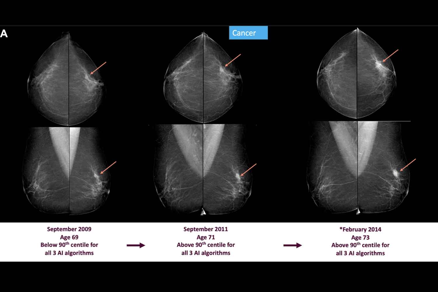
A mammogram can remain on file for years, silently, without anyone coming back to it. Then an algorithm arrives, rereads it after some time and finds something that had remained untraceable at the time: a higher score, a small anomaly, a signal still too weak to immediately change a diagnosis, interesting enough to ask for more attention.
This is the most concrete part of the study published on Radiology: Three commercial artificial intelligence systems applied to previously performed mammograms showed higher scores in people who would receive a breast cancer diagnosis in subsequent years. The work used the Swedish database Validation of Artificial Intelligence for Breast Imagingwith data collected in four regions of Sweden between January 2008 and April 2019. In total, 88,963 mammograms covering 31,394 people were analysed.
The signal before the diagnosis
The researchers looked back, up to ten years before the diagnosis, using three AI-CAD systems, i.e. computer-assisted detection support tools designed for mammography. In the analyzed sample, enriched to study many oncological cases, 12,072 people had then received a diagnosis of breast cancer. The question was very practical: that software would have assigned different scores to previous mammograms, compared to the images of those who would have remained without a cancer diagnosis.
The answer, with all the necessary precautions, goes in the direction of yes. At a specificity of 90%, i.e. with a threshold designed to distinguish positive cases from negative ones with good precision, the AI reported between 19.0% and 19.7% of future cancers already six years before the recorded diagnosis. Four years earlier the share rose to 25.2%, two years earlier it reached 39.3%. Ten years earlier, the signal was weaker, but present in a range between 12.7% and 17.0%.
The strongest data, therefore, concerns the approximately 20% of breast tumors that appear to leave traces on mammographic images that can be read by artificial intelligence already around six years before diagnosis. Subtle traces, often out of reach of the human eye at that moment, or too ambiguous to activate an ordinary clinical pathway.
One more lens
The Swedish system helps to understand the context. The national program invites women between 40 and 74 to have a mammogram every two years, with the reading traditionally carried out by two radiologists. During the period studied, AI scores were available only for research: radiologists reading the exams then worked without that support.
This changes the way you read the results a lot. Here the artificial intelligence looks at images from the past already knowing, through the data, who would have developed a tumor. The study shows potential: using AI scores over time to identify who may need more vigilance, perhaps with additional tests such as an MRI, especially as the radiological profile begins to change from one screening to another.
Prudence remains necessary. The authors themselves recall the limitations of the work: it is a retrospective study, with a dataset built for research; early identification is defined as “potential”; a complete analysis of the exact localization of the future tumor in the images is lacking; and it remains to be understood how those signals can become clinical decisions without increasing too much anxiety, recalls, costs and unnecessary tests.
What changes in screening
The most interesting idea concerns personalized screening. Today mammography mainly follows age, calendar and known risk factors. Mature use of AI could add an extra layer: observe how scores change over time, not limit yourself to the single image. An isolated score can fluctuate, create doubts, generate false alarms. Two consecutive signals, or a sharp increase between screenings, could become more useful in deciding who to screen best.
In Italy, organized mammography screening includes a mammogram every two years for women between 50 and 69 years of age, with possible regional extensions also to the 45-49 and 70-74 age groups. Translated into daily practice, this research alone does not change the indications for patients, but it indicates a very concrete direction for public programs: understanding whether AI can help better choose who to recall, who to follow more closely and who to direct towards additional tests.
Sweden itself, moreover, is already one of the most observed laboratories in this field. Another large Swedish study on the use of AI in mammography screening, the MASAI trial, indicated a reduction in interval cancers and a lightening of the reading burden for radiologists, keeping human control within the path. Here too the direction appears clear: AI works best when it remains a clinical tool, not an automatic shortcut.
The delicate step will be to bring this data out of the archives and into secure protocols. We need prospective studies, continuous monitoring, evaluations of costs, false positives, access to additional tests and psychological impact on the people recalled. An algorithm that sees something earlier can be valuable. Used incorrectly, it can multiply recalls, unnecessary tests and anxiety.
You might also be interested in:
